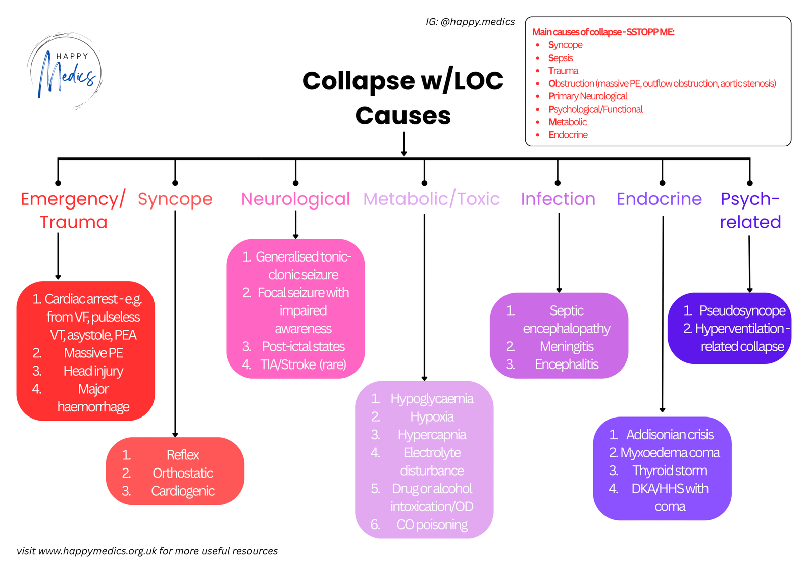
Diagnostic Reasoning: Collapse with Loss of Consciousness Framework

The Pattern Recognition Trap
A 23-year-old medical student loses consciousness this morning after entering his anatomy lab for the first time. He collapses to the floor. When he regains consciousness seconds later, he’s embarrassed but not confused. His instructor says he was unconscious for only a few seconds.
Your System 1 brain sees young patient, emotional trigger, brief loss of consciousness and thinks: classic vasovagal syncope. Reassure and discharge.
But before you anchor on that comfortable diagnosis, there are three questions you need to ask systematically for any patient whohas collapsed: Was there loss of consciousness? If yes, was it syncope? If syncope, what type: cardiac, reflex, or orthostatic?
These three questions form a framework that prevents you from missing the life-threatening causes of collapse, particularly cardiac syncope in young patients with conditions like hypertrophic cardiomyopathy or long QT syndrome.
The Risk Stratification Problem
Transient loss of consciousness is common. Most cases are benign vasovagal syncope. But buried within that large group are patients at high risk of sudden cardiac death (e.g. young athletes with undiagnosed hypertrophic cardiomyopathy), patients with channelopathies (e.g. long QT syndrome), and older patients with structural heart disease or dangerous arrhythmias.
The challenge is that cardiac syncope and vasovagal syncope can look identical in the immediate aftermath. Both patients recover completely and spontaneously. The difference is that one group faces substantial risk of sudden death if the underlying cardiac process that caused their syncope becomes sustained rather than brief.
The System 1 Trap
When you see a young person who’s collapsed and recovered, System 1 anchors on age. Young patient plus emotional trigger equals vasovagal syncope. This is reinforced by experience; most young people who faint do have vasovagal syncope.
But this is precisely where dangerous diagnoses get missed. Hypertrophic cardiomyopathy is the most common cause of sudden cardiac death in young athletes. Long QT syndrome can cause life-threatening arrhythmias triggered by emotional stress, exactly the scenario that looks like classic vasovagal syncope. Both conditions present in children and young adults.
The reverse trap exists for older patients. System 1 sees an elderly person who collapsed and thinks: mechanical fall, maybe orthostatic hypotension. This anchoring can miss cardiac syncope in older patients with structural heart disease, where the mortality risk is substantial.
The problem is that patients with cardiac syncope recover completely after the event, just like patients with vasovagal syncope. There’s no obvious signal that screams “high-risk cardiac disease” unless you ask the right questions systematically.
The Three Questions Framework for Collapse with Loss of Consciousness
Question 1: Was there loss of consciousness?
Not all collapses involve loss of consciousness. Mechanical falls, drop attacks, and some neurological causes present with collapse but preserved awareness. These have different differentials and don’t require the same urgent cardiac evaluation.
Question 2: Was it syncope?
Syncope is a specific subset of transient loss of consciousness caused by transient global cerebral hypoperfusion. Because syncope results from inadequate blood flow to the brain, it has three defining characteristics:
- Abrupt in onset (cerebral hypoperfusion causes immediate loss of consciousness)
- Brief in duration (blood flow must restore quickly or the patient would die)
- Complete spontaneous recovery (when blood flow returns, consciousness recovers fully)
A useful question: “What’s the next thing you remember after losing consciousness?” Any significant confusion lasting more than a minute or two suggests a non-syncopal cause (particularly postictal period from a seizure).
- Non-syncopal causes include:
- Seizures (tonic-clonic activity, incontinence, prolonged confusion)
- Hypoglycaemia (doesn’t recover without intervention)
- Subarachnoid haemorrhage (severe headache, focal neurology)
- Intoxication, trauma, cerebrovascular disease
If the episode was abrupt, brief, with complete spontaneous recovery, it’s syncope.
Question 3: Cardiac, reflex, or orthostatic syncope?
This is the most critical step because it identifies patients with cardiac syncope who are at substantially increased risk for sudden death.
Cardiac syncope is caused by either an electrical problem (i.e. arrhythmia) or a structural problem (e.g. aortic stenosis, hypertrophic cardiomyopathy etc.). These patients need urgent evaluation because the same process that caused brief syncope could become sustained and cause sudden death.
Reflex syncope (including vasovagal) occurs when inappropriate cardiovascular reflexes produce hypotension through bradycardia, vasodilatation, or both. Triggered by prolonged standing, emotional stress, pain, or situational factors. This is the most common type of syncope (20-33% of cases) and generally benign.
Orthostatic syncope occurs when there’s inadequate vasoconstrictor response to standing. Due to volume depletion, medications (diuretics, alpha-blockers, vasodilators), or autonomic failure.
Red Flags for Cardiac Syncope
Certain features should immediately raise concern for cardiac syncope and prompt admission for investigation:
Historical Red Flags:
- Prior cardiac disease (heart failure, coronary artery disease, valvular disease)
- Syncope while supine, sitting, or during exercise (reflex and orthostatic syncope happen when standing)
- Associated chest pain, palpitations, or shortness of breath
- Family history of sudden cardiac death
- Age >60 years
Physical Exam Red Flags:
- Abnormal rhythm, significant murmur (particularly if increases on standing – suggests HCM)
- Gallop rhythm, JVD, lung crackles, significant oedema
ECG Red Flags:
- Sinus bradycardia (<35 bpm) or pauses (>3 seconds)
- Second- or third-degree heart block
- Bundle branch block
- Ischaemic changes
- LVH or RVH
- Long QT (QTc >450 ms males, >460 ms females) or short QT
- Pre-excitation (e.g. delta waves indicating WPW syndrome)
- Arrhythmias
Any of these features means cardiac syncope is possible. These patient would need admission for telemetry, echocardiogram, and further investigation.
It’s also useful to suspect cardiac syncope in patients whose presentation fits neither orthostatic nor reflex syncope.
Features Suggesting Vasovagal Syncope
While no single finding is highly sensitive, certain features substantially increase the likelihood:
Triggers:
- Prolonged standing (LR 9.0)
- Occurring during injection or venepuncture (LR 7.0)
- Abdominal discomfort prior to syncope (LR 8.0)
- Warm environment, lack of food, acute pain or fear
- After exercise (venous pooling)
Prodromal Symptoms:
- Nausea, warmth, diaphoresis, lightheadedness
Context:
- Young patient (<40 years), clear emotional or orthostatic precipitant, normal ECG, no cardiac disease history, no red flags
Orthostatic Syncope
Suggested by syncope immediately upon standing, history of volume depletion, or medications causing orthostatic hypotension.
Orthostatic vital signs: Measure BP and pulse immediately on standing and at 3 minutes. Positive if >30 bpm pulse increase, >20 mmHg systolic BP drop, or >10 mmHg diastolic BP drop, plus symptoms.
The Framework in Action
Back to your 23-year-old medical student who collapsed in the anatomy lab.
Question 1: Was there loss of consciousness?
Yes – he was unresponsive and has no memory of the event.
Question 2: Was it syncope?
Abrupt onset, brief duration (seconds), complete spontaneous recovery with no confusion. This is syncope.
Question 3: Cardiac, reflex, or orthostatic syncope?
Check for cardiac red flags:
- Position: Standing (makes reflex more likely)
- Triggers: Strong emotional trigger (viewing cadaver)
- Prodrome: Queasy, warm, diaphoretic (classic vasovagal)
- Symptoms: No chest pain, palpitations, dyspnoea
- History: No cardiac disease, exercises vigorously without symptoms
- Family history: No sudden cardiac deaths
- Exam: Normal BP and pulse, no orthostatic change, normal cardiac exam
- ECG: Critical – must be done in every syncopal patient
- If his ECG shows:
- Normal ECG: Combined with classic vasovagal trigger and prodrome, no cardiac history and a normal exam, then this is vasovagal syncope. Reassure and discharge.
- LVH or repolarisation changes: Raises concern for hypertrophic cardiomyopathy despite vasovagal-appearing presentation. Needs echocardiogram.
- Long QT: Identifies channelopathy causing life-threatening arrhythmias triggered by emotional stress. Needs urgent cardiology referral.
The framework ensures you don’t anchor on “young patient with emotional trigger = benign” without systematically checking for red flags and obtaining an ECG to rule out HCM and long QT syndrome.
Key Actions: What You Must Do
When assessing any patient with collapse and loss of consciousness, these steps are essential:
1. Always confirm loss of consciousness occurred
Ask witnesses and the patient directly. This guides your differential diagnosis.
2. Use the three features to confirm syncope
Abrupt onset, brief duration, complete spontaneous recovery? Ask “What’s the next thing you remember?” to check for prolonged confusion suggesting seizure.
3. Obtain thorough history focusing on cardiac red flags
- Position when syncope occurred
- Triggers, prodromal symptoms
- Associated symptoms (chest pain, palpitations, dyspnoea)
- Volume loss, medications
- Past cardiac history
- Family history of sudden death
4. Perform detailed physical examination
- Vital signs including orthostatic BP (immediately and at 3 minutes)
- Complete cardiac exam for murmurs (particularly if increase on standing), gallops, abnormal rhythms
- Check for heart failure signs
5. Get an ECG in every syncopal patient
Non-negotiable. Look for rhythm abnormalities, conduction disease, ischaemia, LVH, long or short QT, preexcitation, and any abnormalities. An abnormal ECG is a red flag for cardiac syncope.
6. Admit patients with any red flags for cardiac syncope
- Prior cardiac disease
- Syncope while sitting/supine/during exertion
- Associated cardiac symptoms
- Age >60
- Family history of sudden death
- Abnormal cardiac exam
- Abnormal ECG
7. In young patients with apparently typical vasovagal syncope, don’t discharge until ECG is reviewed
HCM and long QT present in young people and can be triggered by emotional stress. The ECG may be your only clue: LVH, repolarisation changes, prolonged QT, or prominent Q waves should prompt investigation even if the history seems classically vasovagal.
8. Recognise that vasovagal syncope patients need reassurance and education
Young patients with clear triggers, typical prodrome, normal exam, and normal ECG don’t need extensive investigation but do need counseling about avoiding triggers and lying down if prodromal symptoms occur. Counterpressure maneuvers (hand grip, leg crossing, squatting) can abort episodes.
Three Things to Remember
✓ Syncope has three defining features – abrupt onset, brief duration, complete spontaneous recovery. If all three aren’t present, consider nonsyncopal causes like seizures or hypoglycaemia.
✓ Never assume young age means benign vasovagal syncope without checking for red flags – hypertrophic cardiomyopathy and long QT syndrome cause sudden death in young people. Always obtain an ECG. LVH or prolonged QT requires investigation even with a “classic” vasovagal presentation.
✓ Cardiac syncope is the must-not-miss diagnosis – syncope while sitting/supine/during exertion, associated cardiac symptoms, prior heart disease, age >60, abnormal exam, or abnormal ECG all warrant admission. These patients are at substantial risk of sudden death.
About the Author:
Dr Ahmed Kazie is a resident doctor in Acute Medicine and founder of Happy Medics. He teaches systematic diagnostic reasoning through clinical frameworks, helping other resident doctors build confidence in managing acute presentations.
Instagram | Subscribe to Newsletter | Join the AMC Waitlist
